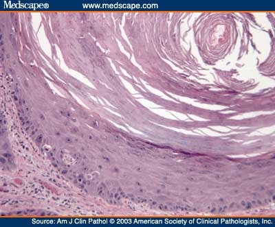
Clinical: epidermal inclusion cyst (follicular cyst)

This photo shows a large epithelial inclusion cysts.

This photograph shows an epithelial inclusion cyst of the lower eyelid.

ISPUB - Epidermal Inclusion Cyst Presenting as a Large Submental Mass

ISPUB - Epidermal Inclusion Cyst Presenting as a Large Submental Mass

Epidermal inclusion cyst-like areas. This nest was located in the deep

Figure 2: Epidermal inclusion cyst of the finger

Epidermal inclusion cyst, skin over breast

Epidermal inclusion cyst

Epidermal inclusion cyst

Epidermal Inclusion Cyst: eMedicine Dermatology

Histopathology Skin--Epidermal inclusion cyst

This is an epithelial inclusion cyst and it is yellow because it is filled

Epidermal inclusion cyst

Epithelial Inclusion Cyst

Epidermal inclusion cyst

Surgial treatment of an iris epithelial inclusion cyst

ISPUB - Infected epidermal inclusion cyst mimicking Marjolins ulcer in a

Epidermal inclusion cyst on the penis of a 55 year old man.

Epidermal inclusion cyst on the penis of a 55 year old man.


No comments:
Post a Comment